Sunday, January 30, 2011

Fun-a-day 30: Blood draws


I'm afraid that the only contact with the medical lab that most people have, is to have their blood drawn for a test.  No one likes needles, including most med techs, and our phlebotomists, who do the majority of the hospital lab's blood draws.  But we do like talking to our patients, even if we do have to stick needles into them.

I hope I've showed you that there's so much more to the lab than a needle!  That said, the blood collection is a very critical part of a lab blood test.  If it's not collected properly, the results can be of no use to anyone, leading to a recollection, which we all hate to do.  Certain tests require the patient to fast, certain tests require other medication or diet restrictions.  Certainly proper labeling is critical and  proper hygiene is very important to protect both the patient and the person who draws the blood.

Safety is certainly another concern, as a stick with a dirty needle can at best cause a lot of worry, and at worst cause severe infections.  We med techs wear lab coats, gloves, wash hands, bleach, and sometimes protective eye wear to keep us safe, but sometimes even that doesn't protect from a broken glass test tube, or a needle.

This is my last quilt block for our fun-a-day project.  We're all going to get together Feb. 10th at the art center to see what everyone else has been working on!  I can't wait!  I hope to get all of the blocks sewn together and the quilt bound in the next few days.  I'll post pictures hopefully soon!  I hope you've had as much fun reading as I have sewing and sharing!

Saturday, January 29, 2011

Fun-a-day 29: Trichomonas


(Oh no she DIDN'T!).  Yes I did!  I debated on whether or not to include this STD / parasite, but I'm afraid it's just another one of those things that are really neat to see under a microscope, even if they're not so much fun if it's your sample that it's found in!  Tricomonas vaginalis is one of the most common sexually transmitted diseases.  It's caused by a single celled protozoa (much like Giardia), and is also considered a parasite.

In the lab, we'd see it in wet preps from OB and ER, or sometimes we'd also see it in a urine sample.  The organism looks similar to a white blood cell, but once you see it moving, or motile, there's no mistaking what it is.  I found this video of it on youtube if you want to see it in action (hope it works).  Luckily it's a curable (and preventable) disease!

I also spent the last few days getting more kindle covers onto my Etsy shop.  I tried for a long time to get a "button" on my blog here.  I think I might have FINALLY figured it out!!  I'm afraid the computer is much more of a challenge to me than any sewing endeavor!  I can't believe that tomorrow will be my last quilt block!  I plan to spend Monday getting it all sewn together!  Wish me luck!

Friday, January 28, 2011

Fun-a-day 28: Alcohol and toxicology

Week-end nights, like tonight, were often a time that we'd get alcohol and drug screens in the lab.  I frequently worked week-end evenings.  In fact I almost always worked Friday or Saturday evening.  I got off around midnight, and dreaded the drive home, knowing and seeing that there were drunk drivers out there.  Blood alcohol levels is a test ER would order on certain patients.  Frequently a urine drug screen also came with it.  Our ER staff was pretty good at predicting the blood alcohol levels of these patients, and usually could also predict what drugs were "on board".  Marijuana, cocaine, heroin, and methamphetamine were among the tests we did.  We ran tests for many therapeutic drug levels too (prescribed medication).

As you might guess, there are some legal issues involved in drug and alcohol testing.  We often had the sad task of testing newborn babies for illegal drugs.  Thankfully the vast majority were negative.  Drunk driving kills far too many people each year.  Unfortunately we were sometimes asked to draw blood for police to send to their crime lab for legal DUI testing.

I put the chemical symbol for alcohol on my quilt bock, and a few other items to add color.  I hope you enjoy your week-end, responsibly, I might add.

Thursday, January 27, 2011

Fun-a-day 27: Chemistry


Chemistry in the lab isn't a whole lot of fun to look at.   There are pretty colored reagent, but much of the fun chemistry is all highly automated now.  Instead we med techs "drive" these huge robotic automated chemistry analyzers.  It's one of the largest sections in the lab, and incredibly important though, so I figured I'd put it in a quilt block.

In chemistry, we deal not with the cells in the blood, but with the serum, or plasma, which is the liquid part of the blood.  We first must spin the blood down in a centrifuge, then do the testing.  We used to have to separate all of the serum off from the blood cells manually, but now many tubes have a gel layer that separates the blood for us.  Our instruments have even started to have "cap piercers" so we don't' have to manually take the top off of each test tube (and put it back on).  The tests we do in chemistry is everything from electrolytes, like sodium and potassium, to enzymes that measure liver function or heart disease, to lipids, like cholesterol and triglycerides.  The amount of science and technology that goes into these chemistry analyzers is incredible, but it still takes a med tech to make sure the instruments are working well, calibrated, full of reagent, accurate, and that they make sense.  We also have to interpret the results, and call critical lab results to the doctor or nurse.

As with chemistry, there are many parts of the medical lab that are hard to make into a quilt... phlebotomists, who draw the blood, couriers who transport it, secretaries, specimen processors, and other critical support staff, managers, special chemistry, coagulation and serology departments, histology and pathology (which I don't have a lot of experience with, but would probably make a great quilt). 

For my quilt top, I "drew" an "SST" tube.  The bottom layer (red) is the cells that gravitate to the bottom when they are spun down in the centrifuge.  The white middle layer is the SST gel that separates the 2 layers.  The top yellow part is plasma, or serum, the liquid part of the blood.  I also drew tests that would normally be part of a basic metabolic panel.  These chemistry tests can be run very fast, and are frequently ordered "STAT".  These few tests can give a doctor a surprising amount of information, which can be critical in emergency situations.  So next time you hear someone order a "STAT" BMP, or chemistry panel on your favorite TV show, you'll know a little bit more about what they're asking for!

Wednesday, January 26, 2011

Fun-a-day 26: Streaking! And WIP Wed.


Sorry, I just couldn't resist!  I was brain storming for ideas today, and I don't know, I guess I stepped back and tried to think of other "circles" in the lab.  Then BAM!  (as Emeril likes to say), it came to me!  One of the "circles" we see the most in the lab is the Petri dish.  In microbiology, we use them a ton!  I didn't regularly work in micro.  Most of the techs who work there are "specialists", even in small labs.  In a smaller lab like where I worked though, most techs regularly do "STAT" (tests ordered to be done immediately) gram stains, and "streak plates".

In a microbiology lab, "streaking" means taking an inoculation loop (the straight line with a circle on the end of it), and streaking it across a petri dish in a way that isolates individual bacteria, that go on to grow out pure colonies that the microbiologist can then identify.  It's a bit complicated, but basically you wipe a sample swap on the dish, which has nutrient-rich growth media on it, spread it out one way, turn the plate a bit, spread it out in another direction, and so on.  On my quilt block I deiced to make the streak lines yellow, then add my interpretation of the bacteria colonies in green.  This is what it looks like for real:


I don't know why, but I LOVED streaking plates.  Maybe it was because it was kind of like drawing.  Maybe it was heating up the loop to glowing orange in our modern Bunsen Burner each time we changed directions of the streak.  Maybe it's just 'cause I'm a bit odd.  But I loved it!

Besides my fun-a-day quilt block, I also spent some time today getting my "New Orleans" art quilt out and finally pin basted it.  I'm trying out "Hobbs batting" for the first time.  My favorite is "Warm and White" up "till now.  What's your favorite?  It's ready to quilt now!  Any suggestions?  I'm linking up with "Freshly Pieced" WIP Wed (yes, I'm really late, AGAIN) to share what we're all workign on today.  Feel free to offer suggestions on how to do the quilting (hint hint).  I'm thinking "simple", but have had others tell me to "quilt the *^&% out of it"! (hee hee).  I've been asked to enter it into our local quilt show.  We're a small town, so it'll be no big thing, but big for me!  Any tips on how to do a "show quilt" are appreciated too, as it'll be my first "show quilt".  And don't worry... I'm a pretty mellow, type B personality, so my aim is more to share than win LOL!

Anyhow, thanks for reading!  I've been really having a lot of fun with this, and am ALMOST sad to be seeing it come to an end in just a few days.  But all good things must eventually come to and end!

Tuesday, January 25, 2011

Fun-a-day 25: Pneumonia


Yep, it's that time of year, cold and flu season!  I've had a sick kid at home the last few days, but it looks like he's on the mend.  Rarely, though, these not-so-fun illnesses can progress to pneumonia, as has happened to another family member of ours in another city.  I hope he feels better soon!  I've had pneumonia once as well, when my kids were both toddlers, and it really can wipe you out for weeks to months! 

There are many causes of pneumonia, but one of the main ones we saw in the lab was caused by a bacteria, Streptococcus pneumoniae.  On a sputum or "bronc lavage" specimen, they appear as gram positive diplococcus, which are the dark dots in pairs on my quilt block.  Since it's a gram stain, the white cells, which are typically quite numerous, appear pink. 

For fun, I drew a "goblet" cell (far left), which we would occasionally see in a bronchial wash specimen, which is a special collection method done in the hospital.  These little cells line the respiratory walls, and have little hairs, or cilia on them that trap foreign particles.  The white cells (on the right) are in there gobbling up the "bad guys", in this case, bacteria, and we do in fact frequently see the bacteria inside of the cell that's just "eaten" them.  So cool!  It still amazes to me, after all of these years, to think (and actually be able to see) that we have these little single cells in our bodies, all constantly working together, each doing their job to keep us healthy.  It's also equally amazing that a tiny virus or bacteria can make us so sick!  I sure hope all of you reading this are healthy and doing well!!

Here's one last picture of my stack of blocks after I trimmed them all up tonight! FUN!

Monday, January 24, 2011

(Not so) Fun-a-day 24: Leukemia


I've been avoiding today's block, as it's probably the cell we med techs dread seeing the most.  But it's also among the most critical things we see under the microscope.  Leukemia is a cancer of the blood, or bone marrow (where the blood is made).  There are many different types, depending on the blood cell line that it originates in.  I "drew" acute lymphoblastic leukemia.  The cells are what we call "blasts", or very early immature lymphocytes.  They have nuclei that are really open and lacy, and very little cytoplasm (the light blue).  There are also typically many, many of them, and they all look very much alike.  Because they multiply unchecked, they can exclude other types of cells.  The white cell count can also skyrocket.

The absolute worst cases are those involving children, especially when we are the first ones to diagnose it.  We hate that call to the doctor or nurse, knowing a patients' life is about to be turned upside down.  Luckily there has been much improvement in the care of leukemia patients, and hopefully that trend will continue.  Unfortunately it's the most common cause of cancer in children, and is much too common in both children and adults.  Among the few dozen blogs I follow, I can think of at least 2 who have family or friends battling this cancer right now.  It's certainly something I personally saw much too often in the lab, as just one case is too many.  I dream of a day where leukemia and all cancer has a permanent cure.

Sunday, January 23, 2011

Fun-a-day 23: Pelger-Huet and my birthday!!


Yep, it's my birthday!  Not only that, but it's my twin brother's birthday as well!  Our birthday had me thinking about genetics.  In a typical hospital lab, there's many many tests that we just don't do, sending them out to a big-city lab.  Genetic testing, is one of many specialties that we didn't do.  Pelger-Huet anomaly is a very rare genetic disorder that causes no symptoms in people who have one copy of the gene (heterozygous), and non-life threatening symptoms in those with 2 copies (homozygous).  But it does cause a striking view under the microscope, as all of the neutrophils only have 2 lobes of their nuclei, instead of the typical 4.  They look like eye glasses.  And of course I see them as "twin" lobes.  I've also seen this in animals too.  It's something I saw only a couple of times.

On my quilt, the Pelger-Huet cells have the bi-lobed dark nuclei, and as usual, I had to throw in some RBC's and platelets to keep them company!

Thanks to my friend, Laura, I'll be eating key lime pie for the first time ever for my birthday.  My hubby's making burgers for the grill as I type!  It'll be as close as we can get to being in sunny Florida!  Maybe we'll even put on some Jimmy Buffet too! Laura has been incredibly busy writing travel guides for Moon Guidebooks, including one that JUST came out for the Florida Keys.  She also has a blog, and this recent post is what got me craving some yummy pie!  My brother (and mom) are both in the Dallas, TX area, so phone calls will have to bring us together today.

Saturday, January 22, 2011

Fun-a-day 22: Spinal Tap and Meningitis


For today's block, I decided to try to "draw" what we'd see on a "hemocytometer" when we look at spinal fluid in a patient with meningitis.  A hemocytometer is used when we want exact counts of cells in a specific amount of liquid.  When we fill the very precise chamber with fluid, using a tiny pipette, only a certain amount of fluid covers the grid lines on the special calibrated glass slide.  It has grid lines that we can use to count and calculate the number we see.  Yes, lots of math!  We not only count spinal fluid cells this way, but also body fluids like pleural or peritoneal.  We also use the hemocytometer to count sperm in the sperm count.

A normal spinal fluid sample would have almost nothing in it.  It's a crystal clear fluid that surrounds the spinal cord and brain.  A spinal tap is very invasive, so we treat the samples in the lab with great care and urgency.  We usually run many tests on it, from cell counts, types, cultures, chemistry, and some are even done at the bedside.  If we do see a significant number of cells, we'll do a differential, letting the doctor know what type they are.  This is important, because once again, if it's a virus, the cells will mostly be lymphocytes.  If it's bacterial meningitis, we'll see mostly neutrophils.  Bacterial meningitis is an incredibly serious illness, and is also contagious.  Thank goodness that we now have some vaccines to help prevent some of the bacterial meningitis, as it's one of the most devastating illnesses we can see in a young child or a young adult.

Once again, this is a "wet prep", so no pretty colors.  Since my husband grew up in Chicago,['; I decided to use some orange thread to go with the blue background (Go Bears!)!!  I also tried using 2 top threads instead of 1.  The little round circles are supposed to be white blood cells.  I'm not too thrilled with the result, or the whole block in general, but I can say I tried something new today!!

Speaking of something new, here's a new ebook cover that I put in my Etsy shop today!  I LOVE this fabric!! Fun!

Friday, January 21, 2011

Fun-a-day 21: Antibodies


Gosh, have I had a busy day!  I made up a few more kindle covers that I still hope to get in my Etsy store tonight, or tomorrow, and I almost finished making up the rest of my fun-a-day quilt blocks.  They're like little canvases, but each still needs to be prepared.  The method I'm using is called "quilt as you go", because that's what you do!  So I have now made up 30 little quilt sandwiches, and 21 are now finished!  All I'll need to do at the end is to connect them all together!  That said, I decided to keep my quilting ridiculously simple tonight!

The symbol stitched on the quilt is that of an antibody.  Antibodies are not only important in our bodies, but in the lab as well.  We can't really see them on a microscope, or at least a light microscope.  They're a protein chain, and are very tiny.  The very ends of an antibody each recognize a very specific "antigen", or foreign protein.  Our bodies make (and remember) an antibody to each virus or bacteria, or perceived "bad guy" that comes along.  An antibody to Hepatitis B is different than an antibody to Influenza.  As with influenza, and colds, and many other things, there is often many different types or strains.

Short story long, scientists have learned how to test for these antibodies.  Not only that, but they've learned how to make these antibodies to use in testing, and indeed many of the tests we do in "special chemistry" in the lab use these in different methods.  I couldn't even begin to list all of the tests where antibodies, are involved.  That's why I decided to use it as a quilt block.  If you've ever had blood drawn to be tested for Hepatitis or HIV, Rubella, or Lyme Disease, we're actually measuring and looking for the antibodies in your blood that shows if you've been exposed to these.  Many tests, such as blood typing, TSH, and sex hormone levels (estrogen, testosterone levels) use manufactured antibodies as a way to test for these levels.  The sophistication of some of these tests, and the instruments that run them are really quite incredible!

I also got nominated by my friend Beth, of Notes ... blog for a Liebster Award,  bestowed upon blogs that could use a boost in followers… (should have less than 300 followers).  If you haven't visited my friend Beth's blog, you really should!  She's a quilter who moved to South Africa about a year ago.  She's also an amazing photographer.  She has some great subject matter to take pictures of!!  She also has helped my via email both with my quilting skills and blogging and message board know-how.  Thanks!

In return, I'm asked to nominate 3-5 other bloggers who could use a boost.  I wish I could nominate all of you, because I really do enjoy everyone's blogs, and know just how much free labor they involve ;-).

Monika of My Sweet Prairie, Kit Lang, of Diva Quilts, and Caroline of Trillium are 3 of many blog that I go to get some inspiration!!

With that, I'm off to bed to read a bit while the wind howls outside.

Thursday, January 20, 2011

Fun-a-day 20: Urine Casts


I have been thinking a bit about kidneys lately.  I'm a big fan of "professional quilter", Mark Lipinski, and have been following his Facebook updates, blog, and Internet radio show, "Creative Mojo".  If you haven't listed to it, you really should (Wed. from 12-2 EST on Toginet radio)!  Lately, he has shared his struggles with a genetic kidney disorder.  It seems that I also have some "kidney issues" on one side of my family, and I also have been trying to make some sense of it.  So, I decided to "draw" urine casts on my block today.  These are generally seen in the urine when a person has kidney problems.  The problems can be many things, including kidney infection, and kidney failure.

On my block, I also put an epithelial cell (skin cells) all the way to the left, which is normal in a urine sample.  Next to it is a "cellular cast".  At the top right is a hyaline cast, which can be seen sometimes in healthy patients.  Below it is a "granular cast", and a "broad waxy cast" is on the bottom.  These are usually only seen in cases of severe kidney disease.  There are other casts as well.  They're formed when an excess of protein is present in the tiny tubes that collect urine.  The protein (which is not normally present in urine) make things "sticky", catching cells and such.  They eventually break free, which is just what a cast is.

On another note, I sold my first ebook case!!!  Whew hew!!!

Just got to add, that I started this post in the afternoon, right before I had to make a fast dinner  for the family (elk steak chunks wrapped in bacon, broccoli and couscous), then take the boys to karate lessons.  I didn't quite finish.  Karate lessons are not just karate lessons!  They are also our "date nights" (DH and I).  We drop them off, then have a blissful kid-free hour at the local coffee house, The Great Pacific (that also sells beer, wine and food)!!  Sometimes we go to a new brew-pub, The Prodigal Son.  It is so totally worth the money we pay for the lessons!  No doubt that my kids will someday become black belts, because we just don't want to give up our date nights!!!  Tonight when I we were driving home from karate lessons, I was talking to the kids about my blog, but I also had to go to the bathroom, so "Becky's bladder" was what the kids heard!!  OMG!  LOL!  Just thought that it was so fitting for today's post!  And it's funny how one thing leads to another!  By the time we got home (only a 4-5 minute drive), I was telling them stories about how my mom spent part of her childhood in a house that had "no indoor plumbing"!  LOL!!  True story!  It's amazing how fast things change!  I love telling my kids family stories like these.  They're so important!

Wednesday, January 19, 2011

Fun-a-day 19: Mononuceosis and atypical lymphocytes


Are you still with me?  I hope so!

Again, I know that mono isn't fun at all, but again, under the microscope it is quite a distinctive illness.  I totally am guessing here, but I believe it was named "mono", because of the abundance of atypical lymphocytes that we see in a blood smear that resemble monocytes.  In any viral disease, the lymphocytes are the heroes.  They multiply rapidly, fighting off the viral infection.  In the process, they appear "reactive" on the slide... the nucleus (purple) gets a bit spread out and lacy, the cytoplasm (light blue) can get quite blue and almost seems to get a bit sticky.  It can look like it clings to the red blood cells on the slide.  This is a very simple "drawing" of what it looks like.  A well-seasoned med tech can spot mono right away.  We always noted the atypical lymphs on the differential, and often called the doctor or nurse to alert them to our suspicion (they usually weren't surprised).  For some reason, these mono infections just look a bit different than most other viral illnesses.  Of course with any viral illness, there's only so much the doctor can do, as antibiotics aren't effective against the virus, but can help if a "secondary" bacterial infection occurs.

Sorry for the bad lighting the last few days.  I've been taking them at night, but better late than never, right?  Speaking of late, I know it's way too late but I'm linking up with "Freshly Pieced" WIP Wed blog again.  We West Coast folks have a bit of a disadvantage time-wise for these linky parties, but maybe there's still a few of us still on the blog-o-spere!

Tuesday, January 18, 2011

Fun-a-day 18: Left Shift, and new Etsy shop!


Just like the bone marrow goes into overdrive making reticulocytes when there's blood loss or anemia, it also goes into overdrive during a bacterial infection, making lots more neutrophils, the white blood cell type primarily responsible for fighting bacterial infections.  They are also kicked out a bit young as the body is trying to recover, and we see this in the nucleus (dark purple).  The nucleus is "band" shaped, and indeed we call these bands in our white cell differential, or "diff".  We look at 100 white cells (under the microscope) in the "diff", and type or sort them, giving the doctor a percentage of each.  If there is a high overall white cell count, and a marked "left shift", the doctor knows that the patient has a severe bacterial infection.  This can be seen in pneumonia, blood poisoning, or appendicitis.  This can be really helpful especially when the patient comes through the ER or has symptoms that can be various things.  Of course I had to put some RBC's and platelets in there too!

But the thing I'm most excited about today is that I got my Etsy store up and running!  It was a bit of a learning curve, but I think I got it.  I hope to add to it and make it "better" as I go along.  I made a few of these ebook cases for family and friends over the holidays, and got such a good response that I'm trying to sell some, as well as iPad covers.  Wish me luck!  Here's the store link: http://www.etsy.com/shop/SolarThreads?ref=pr_shop Since we have 12 solar panels on the top of our house, I decided to call it "Solar Threads".  I'd love any feedback, good or bad if you got a minute!!  Thanks!

Monday, January 17, 2011

Fun-a-day 17: Reticulocytes


Reticulocytes are basically brand new red blood cells that have just entered the circulation.  They are normally about 1% of the total population, but can be increased when the body is working hard to produce new blood cells, as in cases of anemia.  Doctors like to see this response, because it means that the bone marrow is normal and working well.  In cases of a low count, this means that the bone marrow isn't working as it should.  This can be due to several reasons, including bad diet, leukemia, or aplastic anemia.

In the lab, we use a special stain that stains RNA that is present in only very young red blood cells.  The red blood cells all stain a beautiful blue-green.  In my quilt block, the reticulocytes are the cells with little black spots.  You'd normally not see so many "retics" in one "field", but hey, this is a quilt, so I had fun making quite a few of them.  A once time-consuming test, we'd make the smear, stain it, then actually count several fields of 100 cells, and note how many of these were reticulocytes as an averaged percentage.  When I started working in the lab in the early 90's, that's how we did every retic count.  In 2006, when I left, almost all retic counts were then being done on our automated hematology instrument, saving us a lot of time.  As the lab gets more and more automated, we worried as med techs that our jobs will be replaced by instruments.  I don't think that'll ever happen, but certainly our jobs change as we do less hands-on work, and more instrument trouble shooting.

On a totally separate subject, it's Martin Luther King Jr. Day.  I grew up, and attended public schools in the deep South, and was part of the desegregation busing that took place there in the 70's.  I feel fortunate that my parents believed in racial equality and continued to send me to public schools, even as many were being sent to private schools.  I think I learned so much more than math and science by attending the schools that I did.  I hope to continue Martin Luther King Jr.'s dream as I raise my own kids.

Sunday, January 16, 2011

Fun-a-day 16: Bird blood

As I mentioned yesterday, I had zero training in veterinarian lab science in school.  Everything I learned, I learned on the job.  So it was a huge surprise to me to see my first bird blood sample.  All of the red blood cells in birds' circulation have nuclei in them (the black centers).  Apparently all animals except mammals,  have nucleated mature red blood cells.  People, dogs, cats, horses, and even llamas (yes we got those too) have no nucleus in the mature red blood cells after they leave the bone marrow.  We rarely got bird blood in the lab.   Birds don't live too long, in general, and I suspect there just isn't as much a vet can do for a sick bird, so it's not something we saw much at all in the lab.  All of our instruments were designed for humans, so animal blood was a bit of a challenge for us.  But each time we did get a bird sample, it was something of a "come see this!" sample.  We loved seeing new and different things in the lab, and this was no exception.

I didn't even try to "draw" any platelets or white cells, because I just don't remember them at all, but the red cells are pretty fun to look at all by themselves!

Saturday, January 15, 2011

Fun-a-day 15: Veterinary Medicine, Heartworm


One thing medical technology school didn't teach me, and that surprised me when I first started work, was that many medical labs also do veterinary testing.  Animals get sick too, after all, and lab tests, like chemistry panels, and complete blood counts can help the veterinarian diagnose and treat our pets and livestock. 

Heart worm is another mosquito-borne parasite that can be seen in the animal's blood.  It was something that we would occasionally see on a blood smear.  They're quite big, and it doesn't take too much imagination to see how this would cause problems for the animal.  As with other parasitic infections, often there would be an increased number of eosinophils in the blood.  Once again, these are white blood cells that help fight infection, and "specialize" in parasitic infections, as well as allergy response.  The "worm" on the quilt block is a "microfilaria".  The orange cells are the eosinophils.  I put red blood cells and a lymphocyte on it as well for fun.

Surprisingly, dog blood appears very similar to people blood when viewed under a microscope.  One of my favorite "lab party tricks" was to take the "fresh" blood out of the test tube from an infected dog and view it under low power as a wet prep.  These microfilaria often would still be alive and we could watch them move around.  I know, it's a bit warped, but we had to do something in the lab to entertain ourselves...

There are a very few parasites that will form microfilaria in human blood, but luckily I've never seen these.  I think Loa loa (found in Africa) is one of them, but don't quote me on that.  It's been a while since I had parasitology!

Luckily there is prevention and treatment for dogs who get heart worm.  In fact our family pet dog growing up was infected when we brought him home from the pound, and recovered to live a long and happy life.

Friday, January 14, 2011

Fun-a-day 14: Malaria and "twisted" 9-patch quilt


Malaria is a disease that I myself have only really seen once in all of my years in the lab.  It is a parasite that is spread through mosquito bites, and in the United States, mosquito control programs have made malaria almost non-existent here.  But in many tropical and sub-tropical regions of the world, malaria is a real threat, and causes millions of people to get very ill and sometimes die.  Most of the malaria cases diagnosed in the US are from people who have traveled from areas where malaria is present.  Indeed, the one case I "found" in our lab was from such a traveler.  A doctor can also order a malaria smear if they suspect this in their patient.  But malaria is a disease that "waxes and wanes", so a negative smear won't completely rule it out.

The malaria parasite spends part of its life cycle in the human red blood cell.  Other life cycles forms can be seen on a blood smear, but the "ring form" is the most common.  These little tiny rings can be seen inside of the red blood cell.  Luckily there is treatment for malaria, and ways to prevent it.

I made my red blood cells really big so that I could try to make the ring form of malaria easier to see and sew.  Once again, I got a bit frustrated trying to draw something using thread and fabric that is seen on a slide under a light microscope.  But hey, I'm learning a little bit each day!

Speaking of learning, I though I'd share a quick tutorial of a "twisted" 9-patch quilt that I just finished.  I was itching to get it done, as the blocks were finished a few months ago, but put away as I worked on Christmas presents.  I finished the top yesterday, and my husband was home today for lunch and helped me get a good picture of it (even though it was a bit windy).  One thing this quilt taught me is that black isn't all black.  I had some black fabric that I bought in Michigan, and used this all up.  When I bought what I thought was the closest match here in Oregon, only after sewing it all together did I really notice the difference!  Oh well...
A friend of mine told me about this technique about a year ago.  After seeing it on many blogs and in local quilt shows, I decided to make one myself.  It's super-easy and looks great when finished!  When a local store had a sale on fat quarters, I bought 8 of the blue batik fat quarters, then cut out 4, 9 x 9 inch squares from each of them, so I had 32 in all.  I took 2 yards of black fabric and cut out 32, 9 inch squares, and half a yard of the yellow batik, and cut out 8, 9 inch squares.  I sewed them all in 9 patch blocks like this:

Then I cut each of these 9-patch blocks in half both vertically and horizontally, like this:

Now I had 4 smaller blocks.  I twisted them and rearranged them into rows of 5 across, then sewed 6 of these rows together.  I had 2 left over small blocks.  I hope that makes sense.  If you haven't sewed one up yet, do it!  This one was so fun and easy!

Thursday, January 13, 2011

Fun-a-day 13: Sperm Count


I hope I don't offend anyone with this post.  It's truly not my intent, but after doing the fetal hemoglobin block, it's only natural to do a sperm block count next!  So here it is!  Actually a sperm count is only part of a "semen analysis" test.  We also look at the motility (as a percentage) at different times after collection, volume, sperm morphology, and a few other factors.  If infection is present, we certainly note that as well.  When a couple is having problems becoming pregnant, it's usually the first test that a doctor orders, as it is an easy and noninvasive sample to obtain.  Yes, it's embarrassing, but also the rich source of much comedic humor!  It is also usually ordered after a vascectomy, and yes, I have seen sperm in a post-vascectomy sample, but only once that I can think of in my dozen or so years as a med tech.

When we look at the slide, we once again do a wet-prep, so there are no pretty colors, but I just couldn't help but make mine pink and blue, because it is, after all, the sperm that determine if a baby is a boy or girl.  We do the exam on low power, so they look very tiny.  I made mine a bit bigger than what we normally see.  It never ceased to amaze me just how many there are in such a tiny drop (many millions per milliliter).  On a slide, they have no clear "aim", so they swim in all different directions. Once again they really are a wonder to look at under the microscope!

Wednesday, January 12, 2011

Fun-a-day 12: Fetal hemoglobin stain, and WIP


OK  Babies are fun!  One of my favorite "special stains" to do in the lab was a "Kleihauer-Betke Stain" (KLB).  It stains only red blood cells from fetuses or newborn infants (that contain fetal hemoglobin).  The stain is an incredibly vibrant hot pink that's a lot of fun to look at.  We do it on an "Rh negative" blood type mother, when there has been a confirmed fetal-maternal bleed that surpasses the "normal" amount that a regular dose of "Rhogam" would take care of.  Make sense?  I didn't think so.  Rhogam is a drug, created to prevent a potentially horrible condition called "hemolytic disease of the newborn", luckily a disease that has been mostly wiped out due to Rhogam.  This happens when an Rh negative mother carries an Rh positive baby, and is exposed to their blood, usually at birth.  The first baby is fine, but because of the exposure, the mother will develop antibodies, or disease-fighting proteins that target and destroy future Rh positive blood cells, so if she becomes pregnant again with an Rh positive baby, the mothers immune system can literally attack its blood cells.  Rhogam prevents this from happening by binding up all of the Rh positive fetal or baby blood cells before her body can detect them.  I hope that makes sense.

When we do a KLB Stain, we determine by actually counting the number of fetal cells (seen as bright pink) vs the number of maternal (the mom's) cells (see in the black as thin rings, because the stain lyses them).  We give this to blood bank, who then can determine how many additional vials of Rhogam to give to the patient.

I have a personal story about Rhogam.  It was first used in Spring of 1968, just a few months after my twin brother and I were born.  My mom is A neg, I'm O pos, my brother O neg.  After birth I had severe jaundice (which can be a sign of hemolytic disease of the newborn), and was given a complete blood exchange transfusion.  My brother was not.  I know my parents tried years later to have more children, and my mom never got pregnant, so I often wonder if this exposure happened with my mom.

On another note, I'm linking up with "Freshly Pieced" blog's WIP Wed.  I thought I'd show you what else I've been working on.  I made some Kindle covers for gifts for Christmas, and the response was so good that I decided I'd try to see some.  Here's what my kitchen counter looks like today.  You can see my sewing set up in the background... I use our dining room table to sew on.  I do a lot of shuffling things around here!

My "design wall" (ie our bed) has a quilt that I'm itching to finish.  I started it in October, then put it away for after Christmas.  It's a "twisted 9 patch" quilt that was so easy to make, looks great, and all I have to do is sew the blocks and rows together.  Hopefully today, because my bed is also where I fold clothes and I have about 4 loads going today.

Tuesday, January 11, 2011

Fun-a-day 11: Gout


Maybe I should call this "not-so-fun-a-day", because some of these diseases I cover aren't at all fun, including gout.  But once again, it's quite a different story under the microscope.  Gout is caused by needle-shaped uric acid crystals that get into synovial fluid, the normally clear quite thick fluid that surrounds the bones of the joints, like elbows, knees and toes.  All it takes is one look to see why it would cause really severe pain.  I watched my dad go through bouts of gout (really didn't mean to make that rhyme).  The pain was bad enough to send a grown man who rarely went to a doctor, to the ER.

We don't see these very often in the lab, because collecting the sample, is very invasive, and must be done by a doctor.  We use a special polarizing lens (similar to polarized sunglasses), that makes the crystals glow yellow on the vertical plane, blue on the horizontal plane.  Setting up the microscope often is the most time consuming part of the process.  The background is a vibrant fuchsia, and we often see white blood cells in the background, which aren't normal, but are there either from infection or inflammation.  I tried to use purple thread in the background, but am not too entirely happy with the results. 

It's Tuesday, so I'm also linking up with Quilt Story's blog, full of wonderful fabric projects every Tuesday.

And I just have to say that the horrible shootings on Saturday have left me in a real funk.  We recently found out that my husband's aunt and uncle used to live very close to where the shootings took place, and also helped out with Giffords' 2008 campaign, so it's understandably left them quite shaken.  I also think a lot, and remember the deep grief I felt when my own dad was suddenly and unexpectedly taken from our family 2 1/2 years ago (from a pulmonary embolism).  My heart goes out to all of those families who are grieving right now.  I can't even begin to understand what they are going through, but know their pain has to be enormous.

Monday, January 10, 2011

Fun-a-day 10: Urine crystals

We got a lot of the white stuff yesterday, about 4 inches, enough to cause a 2 hour school delay this morning.  With so many tiny snow crystals out there, I thought it'd be fun to put a few urine crystals on my quilt.  A few of you have asked what I'm doing here.  I mentioned in this post that I decided to participate in a January "fun a day" project, through our local community art center (the Pendleton Center for the Arts).  It was started by the Art Clash Collective in Philadelphia as a way to be creative each day for a month, then get together as a local community and share your projects in February.  I think it's such a fantastic idea!

So today I tried to "draw" a few of the most common urine crystals that we see in the lab.  Since urine is such an easy and non-invasive sample to get (ie., pee in a cup), a urinalysis is a very common test ordered by the doctors.  It's amazing what information can be learned from a urinalysis.  It involves 2 parts, a chemical analysis, usually done by a "dipstick", and a microscopic part, looking once again at a "wet prep".  So once again, no pretty colors, but the crystals that we sometimes see are incredibly beautiful.  I'm learning that painting with thread is really a poor substitute for what we see under the microscope.  The light comes from below, and some of theses crystals are 3 dimensional, which refracts the light, making them just glow from within.  If you do a Google image search for "urine crystals", you'll get a better idea of what they look like.  The square "diamond" crystals are calcium oxalate, the rectangular ones are triple phosphate, and the flat ones that look like a Dairy Queen sign are uric acid.  Normal urine is a clear liquid that is sterile until it leaves the body.  The chemical make up can change quite a lot in patients with kidney problems, but also in many other organ diseases and infections.  Of course a urinalysis is also what we use to detect a bladder infection.

And even though I'm not a big sports fan, there's no escaping the fact that the University of Oregon plays a really big national football game tonight (Go Ducks!), so I decided to stitch some green and yellow "O"s into the background!!

Sometimes crystals can be seen in the urine because of kidney stones, but they can also be seen as a normal precipitate that forms as the urine cools.  You'd normally never see more than 1 or 2 different types of crystals in one sample, so this quilt block is just a collection.  Different ones are present in acidic urine, vs alkaline, or basic urine.

Finally, some people have asked how big these are, and how it's coming together.  I made the blocks 8 inches square, with a 5-6 inch diameter (soup bowl-sized) circle that I ironed then appliqued on, using "wonder under" fusible interfacing.  I plan to use binding to join them all together.  I've never done this, so any tips would be much appreciated!  I did some trimming, and this is what it looks like so far.  Enjoy!

Sunday, January 9, 2011

Fun-1-day 9: Fungus among us

This is the quilt block that I sewed yesterday morning.  It has 2 different types of yeast or fungus that can cause problems in our bodies.  On the left, is my attempt at the yeast, "Candida albicans", the main cause of genital  and oral (thrush) yeast infections. "Trichophyton rubrum", on the right is the main cause of skin fungal infections, including ringworm (which is NOT a worm, but does make a ring-shaped patch on the skin), athletes foot, and jock itch.

In the lab, the doctor will order a "KOH prep" to be performed on skin scrapings, or swab to detect these.  KOH, or Potassium Hydroxide, lyses, or dissolves all other cells, and makes the fungal cells easier to see.  It's done as a "wet prep", where the sample is directly examined under the microscope without drying and staining it first. Once again, I used a little creativity, and a little variegated yellow thread for the background.  The yeast and fungus is stitched in grey.

Just like bacteria, yeast and fungus live all around us and in us, normally causing no problems.  In fact, yeast is used to make wonderful food and drinks, like bread,  blue cheese, beer and wine.  This is NOT the same yeast that cause infection though, and it concerns me that when I searched yeast on Google tons of information came up on "yeast free diets", and the like.  Just like this blog, you shouldn't believe everything you read on the internet!

Yeast and fungal infections can be a particular concern in immunocompromised patients.  These patients have a weakened immune or disease fighting system because of cancer treatment, or diseases like AIDS, which affect the immune system.  Yeast and fungi are superb at taking advantage of the change in the immune system, and can rapidly multiply and cause symptoms in large numbers.  In the gut, a course of antibiotics will kill off all the competition for food by killing bacteria, thus allowing the yeast to rapidly multiply, causing yeast infections.  Once again, "icky" subjects, that become beautiful under a microscope...


Saturday, January 8, 2011

Fun-a-day 8: Blood bank

I had already finished my art clash collective Fun'a'day quilt block this morning when we heard on the radio about the tragic mass shooting today in Tuscon, Arizona, at US Rep Gabrielle Giffords public meeting.  Needless to say, we're stunned and horrified.  My  thoughts and prayers go out to Representative Giffords' family and the families of all others involved.  We have family in Tuscon, my husband's aunt and uncle, the recipient of the 50th anniversary quilt that I recently blogged about.  So we are also concerned about them.

As a former medical technologist, my thoughts immediately went to those in the hospital labs all over the city who work in blood bank, including those at the University Medical Center in Tucson.  Also as a former med tech, I have been directly involved in too many traumas to count.  My first 2 jobs were in small hospitals, where I was routinely called to traumas in the emergency room to draw, or collect blood.  I have also worked in blood bank when these traumas came in.  I know first hand what the hospital workers, not just doctors and nurses, but many other departments go through emotionally when they are facing such a situation.  The only thing that makes it bearable is the incredible teamwork that happens and comes together while we all try to save a life.  It's really something that can not be explained.  But my thoughts and prayers go out to all of the many hospital workers, including medical technologists who are working in these cases today.  In situations like this, the teamwork extends to all of the hospitals in the whole area, as well as police and EMT's, firefighters and even people you wouldn't think of, like cab drivers who pick up supplies for us, or couriers who deliver needed blood products and supplies to the hospital.

I wasn't really planning on doing a "blood bank" quilt block, as my plan was originally to do just slides and stains that we see under the microscope (thus the circles), but I felt compelled to stitch together this unit of O negative blood to add to the project today.

People with O negative are called "universal donors", because O negative blood can be given to anyone with no adverse reactions.  If you transfuse the "wrong" type of blood into a patient, it can be fatal.  In a trauma case, we don't have the time to "type and cross match" the blood, so the doctors order O negative blood initially to give to the patient.  The blood bank in the lab will draw a blood sample before any blood has been given to the patient, then determine the blood type ( A, B, AB, or O), and the Rh factor (either positive or negative).    In a cross match, we take blood from the donated unit (we use the little segments that trail off of the unit on the left), and mix, or "cross" it with the potential patient or recipient to see if it's compatible.  In a trauma, as soon as we know the patient's blood type, we switch to "type specific" blood.  That's cross matched too.  It's critical that we keep an adequate supply of O negative blood on hand in case of traumas, but also because people who are O negative can only receive O negative blood.  People who are AB positive are the universal recipient, and can be transfused with blood from anyone with no ill effects.

Forgive me if I don't make much sense today.  The TV has been on all day, and it's hard to concentrate.  But I just wanted to continue this project in a way that helps me make sense of what happened today.

Friday, January 7, 2011

Fun-a-day 7: Gram Stain


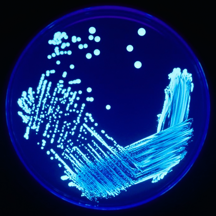
Today I decided to do my "interpretation" of a microbiology Gram Stain.  Gram Stains are one of the most common stains in the medical lab, and are used to differentiate different types of bacteria.  Almost all bacteria are classifies as "gram positive" (which stain purple), or "gram negative", which stain pink.  Furthermore, most bacteria that cause disease in humans are either "rods" (long and thin, like a sausage), or "cocci", (which are little dots or spheres).

I think it was a gram stain that I performed many years ago that was part of my inspiration for my fun-a-day project.  I've been a stay-at-home mom for the last 4 years, but recent events have had me spending a great deal of time thinking about gram stains.

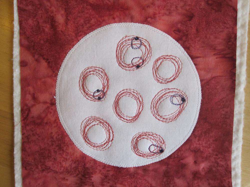
You really can't tell from looking at a gram stain what the bacteria is.  There are so many that can cause infections in people.  We have them living all over and in our bodies, and some of these critter can be "good guys" one day, "bad guys" the next.  Staphylococcus aureus is one of these, seen as purple (gram positive) clusters of cocci.  They remind me of grape clusters.  They normally live happily on the skin, but can infect wounds and other body parts.  The chain of purple dots is Streptococcus bacteria.  There are multiple types of "Strep", and they can cause Strep throat, as well as many other infections.  They remind me of a string of beads.  The big long purple rods are my "take" on Clostridium bacteria.  Different species of Clostridium can cause botulism and tetanus, among other illnesses.  As you may know, they can be found in the soil, and can be very hardy organisms.

Gram negative (pink) bacteria include the rods like E.coli (Escherichia coli) and Salmonella (2 groups at the top), and cocci, like Neisseria, who like to hang out in pairs (far right).  E.Coli is another one of those good guy/bad guy bacteria that normally live quite happily in our lower intestine, but can also cause bladder infections (other bacteria can too), among other things.  A particular strain of E.coli, the 0157:H7 strain, is the one that can cause severe life-threatening illness in undercooked ground beef.  You probably know Salmonella as a cause of "food poisoning".  Different species of Neisseria can cause gonorrhea and meningitis.

The last bacteria, lower center, is one that I myself have never seen, that of Vibrio Cholerae, the cause of Cholera.  This is what's causing so much misery to those in Haiti right now.  It's a curved (like a comma) gram negative rod.

Gram stains can not be used as a sure way to identify the exact species of bacteria that is making someone sick, but can help the doctors know which antibiotic to use, as many are targeted towards these broad classifications.  It gives both the doctor and the microbiologist a really good idea of what the bacteria is and additional tests (culture and sensitivity) can further identify the organism and determine further treatment.

Once again, this is all for fun and shouldn't be used in any other way.  You really shouldn't believe everything you read on the internet anyway, right??  But hope you're having as much fun reading it as I am sewing and writing it!  As a stay at home mom, it's a good way to keep my noodle working, although lately middle school algebra has been using those neurons too :-).

Thursday, January 6, 2011

Fun-a-day 6: Giardia

Again, Giardia isn't such a fun thing to have, but it sure is a really fun (a day) thing to look at under a microscope!  They look like little faces (feces faces ?!?! oh I'm going to get in trouble for that)  looking at you, almost cartoon-ish.

Giardia is the most common parasitic infection of people in the United States, as well as much of the world.  People become infected by drinking contaminated water, such as from streams and lakes.  Beavers are a common carrier of the parasite, so anywhere a beaver may live is likely to have Giardia.  Dogs and other animals can also become infected, and can spread the disease through their feces, or stool (so can people).

Sorry, the medical technologist's world is full of all sorts of "gross" things, as we regularly examine stool, urine, blood, sputum, and various other bodily fluids.  And indeed, Giardia is usually found during an "O and P", or Ova and Parasite stool test.

Stool certainly looks much better on a microscope under 100 power, but I decided to take a little creative liberty and just used a little pretty green thread as a background.  I was going to fill in the giardia with a pastel purple, but when I looked through all of my thread, I realized that I not only don't have pastel purple, but I do not own a single spool of any pastel thread!!  Hmmm  learned something about myself today...

Also, I have a few fun pictures to share with you too.  The local paper mentioned that there's now a huge elk herd that's come down very close to town with all the cold weather we've had.  I took a trip out to see them, and here's a few pictures the kids and I took yesterday!  (Beth, this is where your "manly camera" would have come in handy LOL).  Enjoy!


Wednesday, January 5, 2011

Fun-a-day 5: "Poik"

Today I stitched up my version of poikilocytosis (such a fun word), or abnormal shapes of red blood cell (RBC).  In normal blood, the red cells are all pretty much the same size and shape. In the middle is a normal RBC.  Various medical conditions can cause the red blood cells to become abnormally shaped.  When a medical technologist looks at the blood smear, he or she will rate the amount of "poik", and list all of the abnormal shapes he or she sees, sometimes rating the amount too.  Certain abnormalities can indicate a medical condition.  I'll list the most common medical conditions in parentheses, but once again, I'm not a doctor, so keep that in mind.  Also, some have multiple causes.  There are also "medical" terms for these, and "common" terms, which I interchange.

At the top, working around clockwise, the little cell at "12" is a sperocyte (auto-immune hemolytic anemia), sickle cell (sickle cell anemia), target cell (seen in many anemias, including liver failure), acanthocyte (severe liver disease), stomatocyte (hereditary stomatocytosis), elliptocyte (also common in many types of anemias, including iron deficiency), schistocytes (red blood cell fragments, may be seen in "DIC", or disseminated intravascular coagulation), teardrop cell (severe iron deficiency), and lastly, a nucleated red blood cell (these are immature red blood cells that are normally found in the bone marrow, but in severe anemia when the body is trying hard to produce more RBC's FAST, they can sometimes be seen in the blood).

Phew!  I hope I spelled all of these correctly!  I think it took me longer to write the blog than to sew them!!

One thing I'm learning about "thread painting", is that I really need one of those "open toe" feet on my sewing machine so I can clearly see what I'm doing!  Also trying to fill in an area with dense thread sewing is hard!  My machine doesn't like it either!  Hmmm maybe that's why they make embroidery machines.  But I'm SO not going there!  And it's Wednesday, so I'm linking up with "Freshly Pieced" blog for WIL Wednesday, since this is indeed a WIP!!  Head over there to see what others are working on!

Oh, and here's a few pictures I've taken the last few mornings on my daily walk with my dog.  We've had a long cold snap, that's produced some amazingly beautiful creations...

Tuesday, January 4, 2011

Fun-a-day 4: Anemia

OK, so maybe anemia isn't so fun, but it can be fun to look at on a blood smear, and fun to paint with thread...

Anemia, is essentially a lack of red blood cells, or a disorder of them.  Sudden anemia can occur after a trauma, such as a car accident, major operation, or childbirth (which can be rather traumatic).  A sudden loss of blood really doesn't show up on a blood smear.  The hospital's instruments detect the blood loss in the CBC.  More commonly though, anemia takes a bit of time, because it's due to diet (as in iron deficiency), genetics (as in sickle cell anemia), or due to various other medical problems.

The most common anemia I saw was iron deficiency, which is very common in women, especially when they're pregnant.  It's something I myself have faced, so I thought I'd do a quilt block of what it looks like on the slide.
The red blood cells in iron deficiency anemia are smaller than normal (microcytic), and have less hemoglobin, so they are paler (hypochromic), and look like thin rings.  Instead of being nice equal sized, they can also start varying in size (anisocytosis), and shape (poikilocytosis - isn't that a FUN word?).

Stay tuned for tomorrow.  I think I'm going to do a "poik" block... all the different abnormal RCB shapes.

And it's Tuesday, so Quilt Story is having their fabric Tuesday link party.  Head over there for some inspiration and fun!

Monday, January 3, 2011

"Eos and Basos"

The last 2 types of white blood cells we have in our body's blood are eosinophils and basophils.  Once again, they fight infection, but the eosinphils, especially, play a big part in fighting parasites, and in allergy response.  The eosinophils are my favorite "cell", as they love the eosin dye, which is a vibrant pink-ish orange that just isn't in a thread color pallet.  These are the 2 least common white cells.

Sunday, January 2, 2011

Fun-a-day 2, more WBC's

Today I sewed a few more white blood cells that we all have in our body.  Along with the red blood cell, and platelets, here are the lymphocyte, on the left, and the monocyte, on the right.  Again, this is my interpretation of "normal".

Briefly, lymphocytes are recognized as a main type of cell that kills viruses, and include T-cells and B-cells.  I like to think of monocytes as a kind of "garbage collector", as among other immune system duties, they're good at "phagocytosing", or eating up infected cells and foreign debris in the body.  Lymphocytes are the second most common white blood cell (WBC), and monocytes the third.

Unfortunately, I just have to add, in this litigious day and age, that I am NOT a current medical professional, this is an art blog post, and in no way should you use any of this information for anything even remotely medical related.  Whew...

On a more positive note, I'm having a really "fun-a-day" time with this, and am excited about doing more of these.  I also am realizing that I need to buy a pair of curved embroidery scissors, as my little straight ones just can't cut the thread close enough to the surface of the fabric.  After changing my spool and bobbin a whole bunch of times already for this project, I'm learning that I'd better get used to that too!!

See ya tomorrow!

Saturday, January 1, 2011

Happy 2011, and quilt world meets lab world...

I hope all of you had a great time ringing in the new year!  Let's all hope that it's a kinder, gentler one for everyone!  We let the kids stay up until midnight, then all went to bed a few minutes later!  They should go to sleep easy tonight, especially since they spent the whole day sledding behind our house!

As mentioned, I'm starting my "fun-a-day" project today!  I've already done a little "prep" work, but today I started in earnest.

I'm calling it "quilt world, meet medical lab world".  Part of my medical technologist world was spent looking at countless  microscope slides of blood and other body fluids.  More often than not, they were stained with vibrant dyes and stains to differentiate cells, organisms and cellular structures.  A prerequisite for medical technologists is a color-blind test.  Even before I started school, we had to be tested, because the ability to see color correctly is essential.  One of my favorite aspects of that job, and of quilting is the variety of colors I got to work with each day.

So, for the next 31 days, I'll try to post my thread and fabric interpretation of different things I saw as a medical technologist, starting with this one I did today:
It's a "normal" blood smear.  The red circles are red blood cells, the tiny purple dots are platelets, and the bigger cell in the middle is the most common white blood cell typically seen, a neutrophil.  By looking at blood smears, part of a "CBC", or complete blood count, we can detect many abnormalities, or diseases.  The red blood cells carry oxygen to all parts of the body.  The platelets help stop bleeding when we are injured, and the white cells help fight infection.

Stay tuned to see what's up my sleeve next.  I promise it will involve a needle, but one sewing thread, not drawing blood!!